|
 |
| Adineta cf rhomboidea; creeping specimen, dorsoventral view. Morphotype with similar rhomboid extension on the rump (arrowheads) like in the morphotypes described here, but the spurs are different (arrow). (1) |
| |
 |
| Adineta cf rhomboidea; specimen from the same sample, slightly compessed by coverslide, dorsoventral view. Another trait that is similar to Adineta rhomboidea is the number of nuclei in the vitellarium: 4 (marked by asterisks). This specimen is infected by fungal parasites (marked by arrowheads), presumably Triacutus sp.(1) |
| |
  |
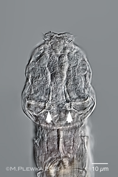
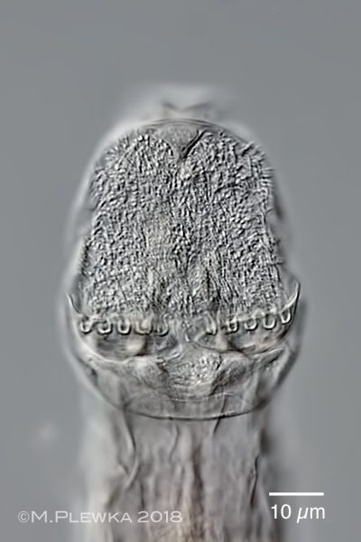
| Adineta cf rhomboidea;; two aspects of the head; dorsoventral view. Left: the arrowheads point to conspicuous light-refracting structures which are actually optical transects of horn-shaped reinforcement structures of the rake blades and which may be the structures that Berzins observed (see also here). Right: ciliary field, focus plane on the rakes. Visible are the U-hooks, the number of which ( NoUH) = 5/5. (2) |
| |
  |
| Adineta cf rhomboidea; foot with spurs; left: crop of the above image; dorsoventral view, sickle-shaped spurs. Right: lateral view while creeping. |
| |
| |
 |
| Adineta cf rhomboidea; another specimen, dorsoventral view; wide rostrum lamella, elevation on rump and spurs are similar to the specimen from 2018. (3) |
| |
 |
| Adineta cf rhomboidea; same specimen, focal plane on the vitellarium with 4 nuclei, which is similar to the Adineta-morphotype from 2014. (3) |
| |
 |
| Adineta cf rhomboidea; gliding, dorsoventral view; specimen from (5). In contrast to the morphotype A. rhomboidea (1) there are no prominent longitudinal folds. 4 nuclei in the viellarium (arrowheads). Wide rostrum lamella. Because the joint between the head pseudosegment (3rd segment) and the rostrum (1st & 2nd pseudosegment) is flexible (see lateral aspect below), the side protrusions of the rostrum lamella have two different outlines depending on the position of the rostrum: leaf-like when the rostrum is inclined to the ventral side or "bird-wing-like" when the rostrum is drawn to the dorsal side by the corresponding retractor muscles. In this image the tip of rostrum lamella is vertical to the slide/ paralell to the optical axis. |
| |
 |
| Adineta cf rhomboidea; same specimen. In this image the tip of the rostrum lamella is directed backwards, so that the outline of the lobes / side protrusions appear leaf-like through the microscope. |
| |
  |
Adineta cf rhomboidea; two images of the posterior part of the same specimen while creeping, focal plane on the spurs. The two images give the impression that the shape of the spurs is different, but:
left image: dorsal view on specimen which is extended.
right image: ventral view on specimen when the toes are attached to the substrate and the toe-bearing foot pseudosegment is perpendicular to the substrate (i.e. cover glass in this case)
This demonstrates that the drawings of different spurs in the literature have to be looked upon as an ambiguous trait for species identification and may have suggested that there are "variations" of the same species in former times.(4) |
| |
 |
| Adineta cf rhomboidea; rake apparatus of semi-macerated specimen; numer of U-hooks (NoUH): 5 in each rake.(4) |
| |
 |
| Adineta cf rhomboidea; ecology: this specimen (lateral view) is infected by fungal parasites (Triacutus cf subcuticularis), some of which are in focus/ visible in the rump. Even when infected this Adineta-species can be very active for another 24h. The two arrowheads mark one of the spores which seems to be partly inside of the head of the rotifer (see below).(4) |
| |
 |
| Adineta cf rhomboidea; ecology: an interesting behavior of several Adineta-species is that the toes of the foot are used to remove particles that stick in the interspace between the ciliary field and the rakes. In this case the rotifer uses the foot to remove a sticky caltrop-like spore. This behavior was observed and filmed for more than 30 minutes, but the rotifer had no success. 4) |
| |
| |
 |
| Adineta cf rhomboidea; another specimen, some days later from the same sample: full of Triacutus-parasites. (5) |
| |
 |
| Adineta cf rhomboidea; at the end of the infection umpteen of Triacutus-spores are ready to leave the corpse of the rotifer (the trophi of Adineta are visible). More on this here >>> (5) |
| |
| |
| |
| |
|
|
| |
| |
| |
| |
| |
| Location: Roscoff, Brittany, France; churchjard, moss (1) St.Pabu, Brittany, France, dune (2); Landildut, Brittany, France; wall in churchyard, (3) |
| Habitat: moss (1); (2); (3) |
| Date: 03.08.2018 (1); 02.08. 2018 (2); 05.08. 2019 (3) |
|